






Anatomy and Physiology of the Vagus Nerve
Overview
The vagus nerve, or cranial nerve X, is one of the most complicated and widely spread nerves in the body. Its name derives from the Latin vagus, meaning “wandering”, reflecting its extensive reach from the brainstem to multiple organs throughout the thorax and abdomen. The vagus acts as the principal channel of parasympathetic communication, regulating heart rate, digestion, and respiratory rhythm and indirectly modulating immune function through neuroendocrine and inflammatory pathways.
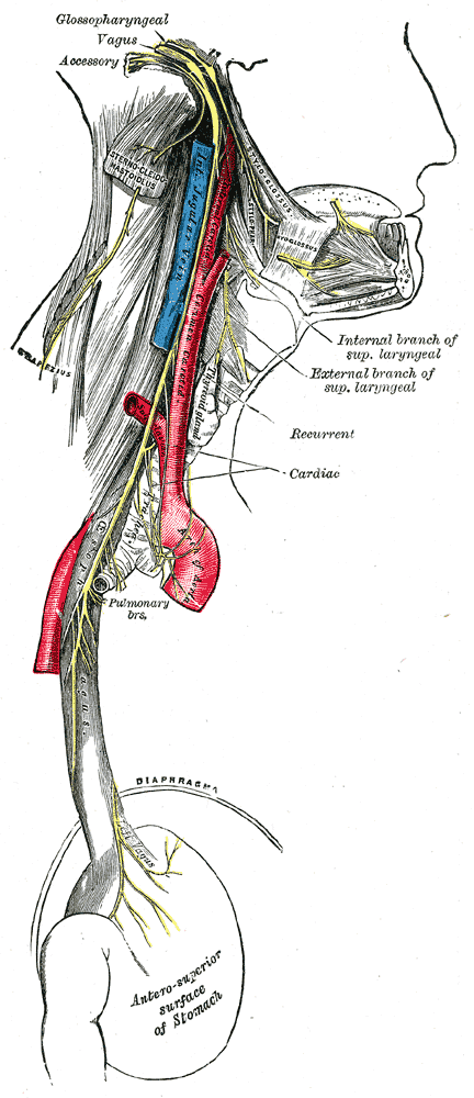
Understanding the structure and signalling pathways of the vagus nerve is essential for appreciating how emotional and physiological states interact. Through its extensive afferent and efferent fibres, the vagus provides continuous two-way communication between the brain and the body, maintaining homeostasis and contributing to the embodied experience of emotion.
Origin and Structure
The vagus nerve originates in the medulla oblongata, where several key nuclei coordinate its activity. These include the nucleus ambiguus, which contributes parasympathetic cardiac control and motor output to the larynx, and the dorsal motor nucleus of the vagus, which provides parasympathetic control to the viscera. Additional input comes from the nucleus tractus solitarius (NTS), which receives sensory information from internal organs and relays it to higher brain centres.
From the brainstem, the vagus exits the skull through the jugular foramen and travels downward within the carotid sheath, alongside the internal jugular vein and carotid artery. Along its course, it branches repeatedly, forming connections with the pharynx, larynx, heart, lungs, and digestive organs. Its fibres are both sensory and motor, providing the structural basis for bidirectional communication between the brain and the viscera.
Nucleus Tractus Solitarius (NTS)
Overview:
The nucleus tractus solitarius (NTS) is a key sensory relay centre located in the medulla oblongata of the brainstem. It serves as the primary destination for visceral afferent signals transmitted via the vagus and glossopharyngeal nerves. These signals include information about cardiovascular, respiratory, and gastrointestinal activity.
Physiological Role:
The NTS integrates sensory input from internal organs and communicates this information to higher brain regions such as the hypothalamus, amygdala, and insular cortex. Through these connections, it helps regulate vital autonomic functions, including blood pressure, heart rate, and respiration. The NTS also plays a role in reflex arcs, such as the baroreceptor reflex, which help maintain stable circulation during changes in posture or activity.
Afferent and Efferent Fibres
Approximately 80% of vagal fibres are afferent, carrying non-painful visceral sensory information from organs to the brain. These fibres report on visceral states such as gut distension, blood pressure, and chemical balance, which contribute to interoception, which in turn shapes emotional awareness. The remaining 20% are efferent, transmitting motor signals from the brain to regulate heart rhythm, respiration, and digestion.
This division illustrates the body’s emphasis on monitoring internal conditions. Through the vagus nerve, the brain constantly receives data on the state of organs, allowing rapid physiological and behavioural adaptation. This mechanism underlies the dynamic relationship between bodily sensation and emotional state: a key area for understanding how IEMT interventions may indirectly influence autonomic balance.
Major Branches of the Vagus Nerve
The vagus gives rise to multiple branches that supply different regions of the body. The major divisions include:
- Pharyngeal and Laryngeal Branches: Control muscles involved in swallowing, speech, and vocal tone. These pathways are essential for communication and social interaction, linking vagal activity to prosody and emotional expression.
- Cardiac Branches: Regulate heart rate through parasympathetic input, slowing cardiac rhythm during rest and promoting recovery after exertion.
- Pulmonary Branches: Supply the lungs, influencing bronchial constriction, respiratory rhythm, and reflexes such as coughing.
- Gastrointestinal Branches: Extend through the oesophagus, stomach, and intestines, coordinating motility and digestive secretions, and indirectly influencing nutrient absorption.
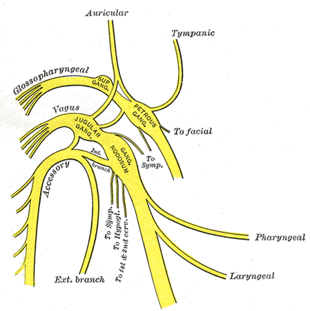
- Auricular Branch (Arnold’s Nerve): A small sensory branch that innervates part of the outer ear. Stimulation of this region has been investigated in non-invasive vagus nerve stimulation research.
Auricular Branch (Arnold’s Nerve)
Overview:
The auricular branch of the vagus nerve, also known as Arnold’s nerve, is a small sensory branch that supplies parts of the external ear, particularly the ear canal and a section of the auricle. It is the only cutaneous branch of the vagus nerve, carrying sensory information from the skin to the brainstem.
Physiological Role:
Sensory signals from the auricular branch travel to the nucleus tractus solitarius within the medulla, linking external tactile stimulation with autonomic regulation. Because this branch connects peripheral sensory input to vagal pathways, gentle stimulation of the ear region has been explored in research on non-invasive vagus nerve stimulation (nVNS). Such stimulation may influence parasympathetic tone and has been studied for potential effects on mood and heart rate regulation.
Functional Integration
The vagus nerve does not operate in isolation but as part of a complex autonomic network that includes sympathetic, parasympathetic, and enteric systems. Its activity is modulated by higher brain structures such as the hypothalamus, insular cortex, and prefrontal regions, which integrate sensory input, emotional context, and cognitive appraisal. This integration allows the vagus to participate in coordinated behavioural responses, including those related to safety, communication, and social engagement.
Vagal signals influence cardiovascular tone, respiratory rhythm, digestive efficiency, and inflammatory regulation. Through mechanisms such as the “cholinergic anti-inflammatory reflex”, vagal efferents can modulate immune activity, reducing excessive inflammation. These functions demonstrate the vagus nerve’s role as both a physiological regulator and a mediator of the mind–body connection.
The Vagus and the Emotional Brain
Vagal afferents reach emotional-processing regions (including the nucleus tractus solitarius, the amygdala, the hypothalamus, and the insular cortex) through ascending brainstem pathways. This circuitry allows emotional experiences to be grounded in bodily sensations - for instance, a quickened heartbeat during fear or a settled gut during calm connection. The vagus thus provides the physiological foundation for the embodied nature of emotion, linking perception, feeling, and autonomic regulation into a coherent whole.
Nucleus Tractus Solitarius (NTS)
Overview:
The nucleus tractus solitarius (NTS) is a cluster of grey matter located in the medulla oblongata of the brainstem. It serves as the primary receiving centre for visceral sensory information carried by the vagus and glossopharyngeal nerves. These signals include data on heart rate, blood pressure, respiratory rhythm, and digestive activity.
Physiological Role:
The NTS integrates incoming afferent information from internal organs and relays it to higher brain regions such as the hypothalamus, amygdala, and insular cortex. This pathway enables the brain to monitor and adjust autonomic output in real time, maintaining stable internal conditions. The NTS also contributes to vital reflexes such as the baroreceptor and chemoreceptor reflexes, which regulate cardiovascular and respiratory function.
In therapeutic settings, awareness of this relationship helps practitioners understand the correlation between alterations in breathing, heart rate, or posture and fluctuations in emotional states. These physiological adjustments are not merely secondary effects; they form part of the broader psychophysiological shifts that accompany emotional change.